Now that I have looked at the basics of what chemokines do in general, I am going to look specifically at the function of CC chemokine receptor 2 and its ligands. Once we know the mechanism of action of this receptor and it’s chemokines and how it functions under normal circumstances we can then go on to look at what its aberrant expression causes and how this can be treated.
Here is a little summary table to describe the properties of CCL2/MCP-1:
Produced By | Monocytes Macrophages Fibroblasts Keratinocytes Vascular Smooth Muscle Cells Endotheleial Cells T cells |
Receptor | CC chemokine Receptor 2 (CCR2) |
Attracts | Monocytes Natural Killer Cells Basophils Eosinophils Dendritic Cells |
Major Effects | Activates macrophages Basophil histamine release Promotes T-helper 2 (TH2) cell immunity |
Adapted from Janeway's Immunobiology by Murphy and Travers (2008).
MCP-1 Structure
There are 4 known MCP chemokines involved in human immunity, MCP1-4. MCP-1 is secreted in response to inflammatory signals in two forms that have molecular weights of 9kDa and 13 kDa. The differences in molecular weights are to do with O-glycosylation but this doesn’t affect their ability to attract monocytes and other immune cells. The residues in the amino terminal are crucial in the functioning of the MCP-1 protein. The mains ones include:
- Residues 1-6: contribute to the chemoattractant ability of MCP-1 (Asp-3 in particular has been noted as a key player)
- Residues 7-10: involved in receptor desensitisation
- Amino Acid at Position 1: important in the secondary structure formation of the protein and therefore the binding of MCP-1 to recpetors
It has been most commonly noted that MCP-1 binds to CCR2 in a dimeric fashion, although monomers of MCP-1 have also been known to cause activation of receptors too (17).
Functions of MCP-1
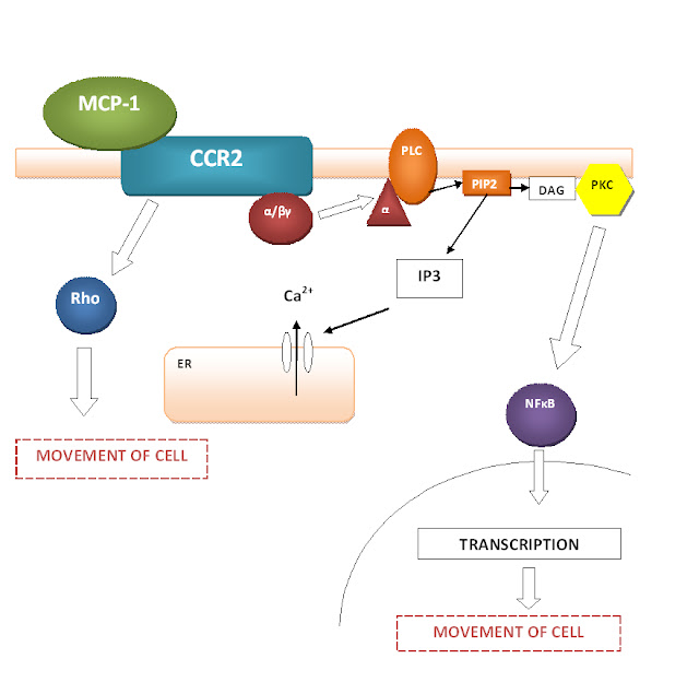
The ability of MCP-1 to cause chemotaxis and the movement of cells is thought to be due to the fact that through the signally pathway, CCR2 activation activates phospholipase C and therefore, eventually increases the concentration of intracellular calcium through IP3 signalling. A schematic diagram is shown to summarise this process along with the other theories of how chemotaxis is enabled: that of NFκB activation and Rho activation.
Adapted from Melgarejo, E. et al. 2009. Monocyte Chemoattractant Protein-1: A Key Mediator in Inflammatory Processes. The International Journal of Biochemistry and Cell Biology 41: 996-1001
MCP-1 Signalling.
Cell motility is enabled through a variety of ways. These include the activation of phospholipase C, causing the cleavage of phosphatidylinositol 4,5-bisphosphate (PIP2). This results in 2 products: diacylglycerol (DAG) and inositol triphosphate (IP3). IP3 can bind to IP3 receptors on the endoplasmic reticulum (ER) to cause calcium increase which is needed for cell movement. DAG causes the activation of protein kinase C (PKC) which can phosphorylate transcription factors like NFκB which is the basis for directional movement of the cell. Another mechanism by which cell movement is enabled is by the activation of Rho family of proteins. These proteins facilitate actin-dependent processes which result in pseudopod formation and membrane-ruffling (17).
Cell motility is enabled through a variety of ways. These include the activation of phospholipase C, causing the cleavage of phosphatidylinositol 4,5-bisphosphate (PIP2). This results in 2 products: diacylglycerol (DAG) and inositol triphosphate (IP3). IP3 can bind to IP3 receptors on the endoplasmic reticulum (ER) to cause calcium increase which is needed for cell movement. DAG causes the activation of protein kinase C (PKC) which can phosphorylate transcription factors like NFκB which is the basis for directional movement of the cell. Another mechanism by which cell movement is enabled is by the activation of Rho family of proteins. These proteins facilitate actin-dependent processes which result in pseudopod formation and membrane-ruffling (17).


No comments:
Post a Comment